 |
| By Dsealy – Own work, CC BY-SA 4.0, Link |
I hope this post is useful especially to student nurses and to those who need to educate patients about ECG/EKGs.😊
Doctors use a test known as an electrocardiogram, also referred to as an ECG or EKG, to measure the heart’s electrical activity and detect any anomalies in one of the most important organs of the body. An ECG makes a recording of the timing and strength of electrical impulses as they flow through the heart. Coming out in a moving strip of paper from the ECG device, the recording is then
read and interpreted by the doctor to see if the heart is working normally.
While it may seem simple to understand, how the test actually works is truly a mind-boggler, especially if one does not know how the heart works in the first place.
The electrical system of the heart
The heart is composed of specialized tissues that are capable of creating electrical signals that cause the cardiac muscles to contract. In every contraction, the heart works by pumping blood out to the lungs in order to absorb oxygen and to deliver it to the cells all over the body.
Particularly, the sinus node, also referred to as sinoatrial node, is the tissue that’s responsible for generating electrical activity throughout the heart. Located in the atrium or the upper right chamber of the heart, the sinus node sends out an electrical impulse in every fraction of a second, traveling through the atrial muscles, which causes them to contract. The impulse then travels through the atrioventricular node in order for it to reach the ventricles of the heart.
As the electrical impulse flows through the organ, the heart normally contracts, or beats, about 60 to 100 times in a minute at rest in most adults.
Understanding the ECG reading
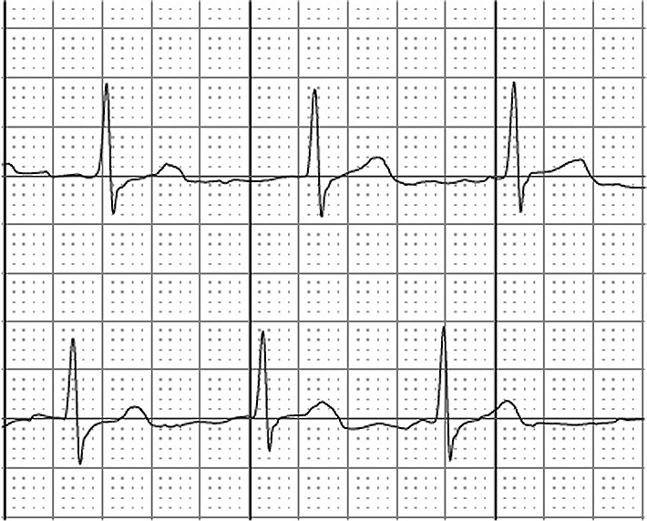
Every activity of the heart can be seen through the electrocardiogram reading, which appears like a line graph with valleys and peaks. These peaks and fluctuations actually represent the “waves” of signals flowing through the heart. The P wave represents the electrical signal sent through the heart’s upper chambers. Meanwhile, the QRS wave indicates the electrical activity in the lower chambers. Lastly, T wave records the heart’s activity as it returns to rest.
 |
| By MoodyGroove at the English language Wikipedia, CC BY-SA 3.0, https://commons.wikimedia.org/w/index.php?curid=2581061 |
Reflected on the ECG print, the size and shape of the waves and the interval between each wave reflect the rate and regularity of the heart activity, which is valuable information for your doctor as it will help diagnose your condition. Aside from shedding light to the heart’s rhythm, the ECG can also determine possible heart muscle damage as well as detect abnormal levels of blood electrolytes, including calcium and potassium.
How it is performed
An electrocardiogram is a painless procedure that often takes only a few minutes. It may be done at the doctor’s office or as a laboratory procedure at the hospital. Since ECG equipment is portable, the test can be carried out almost anywhere. For patients admitted to the hospital, your heart patterns may be constantly monitored by an ECG system.
During the test, the patient may initially be required to shave the chest, arms, and legs to provide a smooth surface to attach the electrodes in the ECG cables. Once the area is cleaned, the patient is then asked to lie on a bed, where several ECG cables and leads are attached to the skin on the chest and on each arm and leg. These cables are connected to the machine that records the patient’s heart activity into print.
The three main types of ECG
An electrocardiogram is required by the doctor depending on three different purposes. The resting ECG is when the doctor wants to know how your heart works while you are at rest. The second type, exercise ECG, is requested if the doctor wants to determine the heart’s reaction to activity. This test may be done while the patient is walking or running on a treadmill. There is also what we call the 24-hour ECG, which monitors the patient’s heartbeat the entire day.
The ECG is an essential test that can detect any damage to the heart so you and your healthcare team can prevent it from getting worse. With the convenience of the test results and your doctor’s intervention, the least you can do is to make changes in your lifestyle and to be more proactive in dealing with your own health.
 window.amznpubstudioTag = “daretodreampr-20”;